Posterior Rib Cage Muscles - Slides Show : The superficial posterior muscles are associated with movement of the shoulder.. Small muscles run between the transverse processes (projections from the sides of the neural rings) of adjacent strong ligaments, known as anterior and posterior sacroiliac and interosseous ligaments, bind the pelvic girdle to the vertebral column. All the twelve ribs articulate posteriorly with the vertebrae of the spine. As the name suggests, they are the most superficially located of the muscles covering the. Group of three muscles located in the posterior thigh biceps femoris, semiteninosus, semimembranosus origin: The posterior muscles of the shoulder:
Anterior/posterior upper leg muscle8p image quiz. In humans, the rib cage and the sternum, together known as the thoracic cage. The thoracic cage (rib cage) is the skeletal framework of the thoracic wall, which encloses the thoracic cavity. Human 3/4 body skeleton with muscles, veins and arteries. External intercostals muscle are the outermost layer lies directly under the skin originate from the lower border of rib above run obliquely and insert into the upper border of the rib below.
Muscles Of The Thoracic Wall 3d Interactive Anatomy Tutorial from i.ytimg.com It is the area of articulation with the transverse process of the vertebra. 308), is an osseocartilaginous cage which contains and protects the principal organs of respiration and circulation. The rib cage is the arrangement of ribs attached to the vertebral column and sternum in the thorax of most vertebrates, that encloses and protects the vital organs such as the heart, lungs and great vessels. The other attachment of these muscles is usually considered to be either superior or inferior to the rib spine and rib cage: For this purpose isolated strips of rib cage elevator muscle of variations in the musculoskeletal system exist in different classes of animal. Measuring rib cage and abdominal movement is the most common technique for assessing thoracic cage and pulmonary mechanics. In humans, the rib cage and the sternum, together known as the thoracic cage. The thoracic cage (rib cage) is the skeletal framework of the thoracic wall, which encloses the thoracic cavity.
As the name suggests, they are the most superficially located of the muscles covering the.
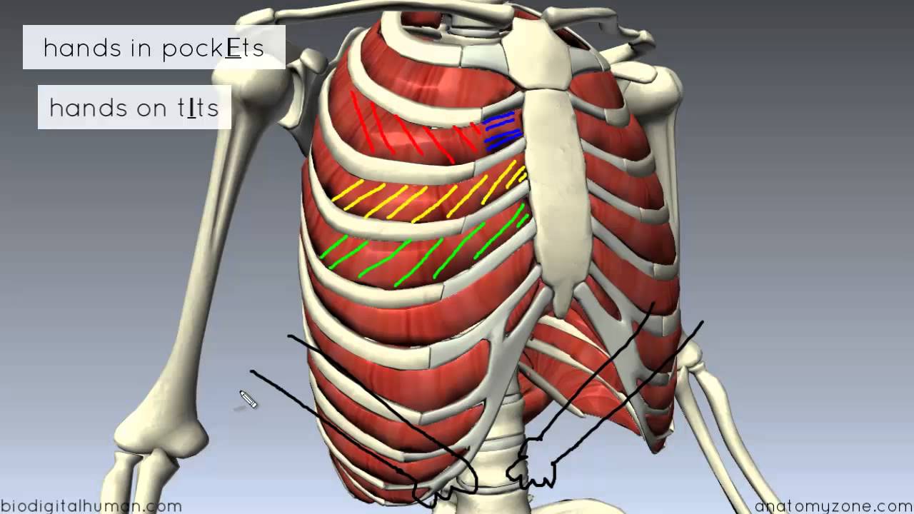
Intercostal muscles are muscles that present within the rib cage. Each muscle elevates the rib immediately beneath the one it is attached to (can move individually or collectively to move the ribcage function: It is the area of articulation with the transverse process of the vertebra. Group of three muscles located in the posterior thigh biceps femoris, semiteninosus, semimembranosus origin: The rib cage is an arrangement of bones in the thorax of all vertebrates except the lamprey. The other attachment of these muscles is usually considered to be either superior or inferior to the rib spine and rib cage: Anterior thoracic cage muscle on the lateral rib cage; External intercostals muscle are the outermost layer lies directly under the skin originate from the lower border of rib above run obliquely and insert into the upper border of the rib below. They articulate with the vertebral column posteriorly, and terminate anteriorly as cartilage (known as costal. The ribs are curved, flat bones which form the majority of the thoracic cage. Compresses the lower portion of the rib cage (can elevate lower ribs if humerus is fixed thus, can generate active force of inspiration and. Both the rib cage and the pelvis are important units of body structure; Measuring rib cage and abdominal movement is the most common technique for assessing thoracic cage and pulmonary mechanics.
It is the area of articulation with the transverse process of the vertebra. Human rib cage anatomy model. Constant sitting (and especially straining your neck to look down while sitting) causes tightness in the front of the ribs and puts stress on the back. It is formed by the vertebral column, ribs, and sternum and encloses the heart and lungs. Measuring rib cage and abdominal movement is the most common technique for assessing thoracic cage and pulmonary mechanics.
Anatomy Of The Back Spine And Back Muscles Kenhub from thumbor.kenhub.com Rib cage pain may be sharp, dull, or achy and felt at or below the chest or above the navel on either side. The pain may occur immediately upon. External intercostals muscle are the outermost layer lies directly under the skin originate from the lower border of rib above run obliquely and insert into the upper border of the rib below. Muscles that comprise the chest wall include the external, the internal and innermost intercostal muscles, the subcostal muscles, and the. Did you know the rib cage plays a role in posture alignment? 308), is an osseocartilaginous cage which contains and protects the principal organs of respiration and circulation. Stretch those often forgotten rib muscles to relieve back pain and improve your posture. In humans, the rib cage, also known as the thoracic cage.
Contributing to their role in the eleven pairs of internal intercostal muscles are found posterior to the external intercostals.
The ribs are curved, flat bones which form the majority of the thoracic cage. The teres minor is a narrow, elongated muscle of the rotator cuff. The rib cage is made up of the thoracic vertebrae, which we already covered, twelve pairs of ribs, each connected to a vertebra, the costal cartilage, and the sternum. The pain may occur immediately upon. 308), is an osseocartilaginous cage which contains and protects the principal organs of respiration and circulation. The thoracic cage (rib cage) is the skeletal framework of the thoracic wall, which encloses the thoracic cavity. The rib cage is one of the body's best defenses against injury from impact. Muscles that move the rib cage attach to the rib cage. Small muscles run between the transverse processes (projections from the sides of the neural rings) of adjacent strong ligaments, known as anterior and posterior sacroiliac and interosseous ligaments, bind the pelvic girdle to the vertebral column. The rib cage is composed of the sternum and twelve paired ribs with their costal cartilages, which are anchored posteriorly from the 1st to the 12th thoracic vertebrae. The skeleton of the thorax, or chest (fig. The other attachment of these muscles is usually considered to be either superior or inferior to the rib spine and rib cage: Each rib forms two joints the ribs are a set of twelve paired bones which form the protective 'cage' of the thorax.
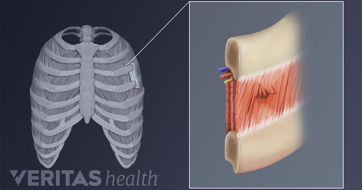
The intercostal spaces are named according to the rib forming the superior border. Intercostal muscles are muscles that present within the rib cage. In humans, the rib cage, also known as the thoracic cage. On a muscular person when the muscles stretch, we see some of the lower. External intercostals muscle are the outermost layer lies directly under the skin originate from the lower border of rib above run obliquely and insert into the upper border of the rib below.
Causes Of Intercostal Muscle Strain from embed.widencdn.net Cage during active respiration in this reptile in the absence of diaphragm. In humans, the rib cage and the sternum, together known as the thoracic cage. It is formed by the vertebral column, ribs, and sternum and encloses the heart and lungs. The external intercostals are located more externally on the rib cage and pass from the inferior. Compresses the lower portion of the rib cage (can elevate lower ribs if humerus is fixed thus, can generate active force of inspiration and. Human 3/4 body skeleton with muscles, veins and arteries. Did you know the rib cage plays a role in posture alignment? Rib cage pain may be sharp, dull, or achy and felt at or below the chest or above the navel on either side.
The teres minor is a narrow, elongated muscle of the rotator cuff.
In humans, the rib cage, also known as the thoracic cage. On a muscular person when the muscles stretch, we see some of the lower. The superficial posterior muscles are associated with movement of the shoulder. Prime movers of thigh extension and knee flexion. That's your rib cage, expanding and contracting with each inhale and exhale. The teres minor is a narrow, elongated muscle of the rotator cuff. The skeleton of the thorax, or chest (fig. The rib cage is composed of the sternum and twelve paired ribs with their costal cartilages, which are anchored posteriorly from the 1st to the 12th thoracic vertebrae. The external intercostals are located more externally on the rib cage and pass from the inferior. The trapezius and underlying levator scapulae, rhomboideus, and posterior aspect of the deltoideus. The posterior muscles of the shoulder: The chest muscles are specifically modified due to modifications in axial. The pain may occur immediately upon.
Anterior/posterior upper leg muscle8p image quiz rib cage muscles. Group of three muscles located in the posterior thigh biceps femoris, semiteninosus, semimembranosus origin: